Bloat
- Thomas W. G. Gibson, BSc, BEd, DVM, DVSc, DACVS
- Feb 16, 2018
- 7 min read
Gastric dilation and volvulus (GDV) is an acute, life-threatening condition that primarily affects large- and giant-breed dogs. Immediate medical and surgical intervention is required to optimize chance of survival.
Etiology and

Pathophysiology:
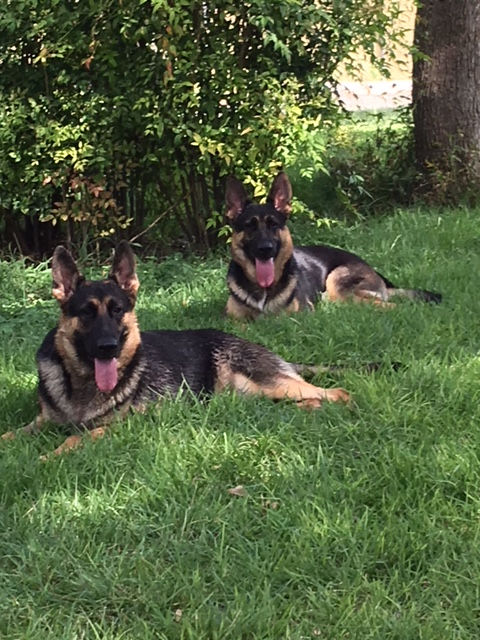
The etiology of GDV is unknown, but several phenotypic and environmental risk factors have been identified for developing GDV. Breeds at risk of GDV include the Great Dane, German Shepherd, Irish Setter, Gordon Setter, Weimaraner, Saint Bernard, Standard Poodle, and Bassett Hound. No sex predisposition exists, and dogs appear to be at increased risk with advancing age. Other reported predisposing factors include lean body condition, deep/narrow thoracic conformation, a first-degree relative with a history of GDV, stress, aggressive or fearful behavior, once daily feeding, dry food, rapid consumption of food, previous splenic disease, and increased gastric ligament laxity.
It is unclear whether dilation or volvulus occurs first during the development of GDV, although it is postulated that volvulus occurs first. Dilation of the stomach results from accumulation of gas and/or fluid, and volvulus prevents the normal release of these contents. During volvulus, the pylorus and duodenum first migrate ventrally and cranially. Viewed from a caudal to cranial direction, the stomach may rotate from 90° to 360° in a clockwise fashion about the distal esophagus. This rotation displaces the pylorus to the left of midline, entrapping the duodenum between the distal esophagus and the stomach. Depending on the degree of volvulus, the spleen may vary in position from the left caudodorsal to the right craniodorsal abdomen. A volvulus of >180° causes occlusion of the distal esophagus.
Gastric dilation and volvulus, dog
Gastric dilation and volvulus, dog. Illustration by Dr. Gheorghe Constantinescu.
After volvulus of the stomach, gas is trapped within this compartment and intragastric pressure rises. Gastric outflow obstruction may be caused by compression of the duodenum by the distending stomach against the body wall, or it may be due to the presence of neoplasia, a gastric foreign body, or pyloric stenosis. Splenic entrapment often accompanies GDV. The progressively distending stomach compromises venous return by compression of the caudal vena cava. Sequestration of blood in the dilated splanchnic, renal, and posterior muscular capillary beds results in portal hypotension, GI tract ischemia, hypovolemia, and systemic hypotension. These factors combine with the loss of fluid in the obstructed stomach and a lack of water intake to produce signs of hypovolemic shock. Dogs are at risk of endotoxemia, hypoxemia, metabolic acidosis, and disseminated intravascular coagulation.
Clinical Findings:
Dogs may present with a history of nonproductive retching, hypersalivation, and restlessness. Acute or progressive abdominal distention may be noted, or the affected dog may be found recumbent and depressed with an enlarged abdomen.
Physical examination findings include an enlarged or tympanic abdomen. Abdominal pain and/or splenomegaly may be appreciated on abdominal palpation. Progression from gastric dilation to volvulus predisposes to hypovolemic shock. Signs of shock are common and can include weak peripheral pulses, tachycardia, prolonged capillary refill time, pale mucous membranes, and dyspnea. An irregular heart rate and pulse deficits indicate the presence of a cardiac arrhythmia. Additionally, the expanding stomach may compress the thoracic cavity and inhibit diaphragmatic movement, leading to respiratory distress.
Diagnosis:
Suspicion of GDV is usually high after considering the history, signalment, and clinical signs. Radiographs help distinguish simple gastric dilation from GDV. The preferred radiographic views for identification of GDV are right lateral and dorsoventral recumbency. Ventrodorsal positioning must be avoided because of the potential for aspiration of gastric contents.
The right lateral radiograph usually reveals a large, distended, gas-filled gastric shadow with the pylorus located dorsal and slightly cranial to the fundus. The gastric shadow is frequently compartmentalized or divided by a soft-tissue “shelf” between the pylorus and fundus. This shelf, or reverse "C” sign, is created by the folding of the pyloric antral wall onto the fundic wall. Splenic enlargement or malposition may be noted on radiographs. Gas within the gastric wall is suggestive of tissue compromise, whereas free gas within the abdomen indicates gastric rupture.
Gastric torsion, dog
Courtesy of Dr. Ronald Green.
PCV, total solids, electrolytes, blood glucose, and serum lactate levels should be evaluated, and blood drawn for a CBC, serum biochemical profile, and coagulation assays. Continuous ECG and blood pressure monitoring are recommended.
Prerenal azotemia is a common finding in animals with GDV and is secondary to systemic hypotension. Increased CK levels may be present due to striated muscle damage, and serum potassium levels may increase subsequent to cell membrane damage. Serum ALT and AST levels may increase secondary to hypoxic damage. Increased lactate is a common finding and is secondary to systemic hypotension and inflammation. Hyperlactatemia (>6 mmol/L) is associated with an increased likelihood of gastric necrosis and the need for partial gastric resection.
Treatment:
Immediate goals in treatment of GDV include restoring circulating volume and gastric decompression. Rapid surgical correction of the volvulus follows initial patient stabilization. Because duration of clinical signs is one of the risk factors of GDV-associated death, it is imperative to recognize and correct this condition immediately.
Correction of hypovolemia is the first treatment priority and is achieved by rapid fluid replacement with one or more large bore (16- to 18-gauge) IV catheters placed in cranial (jugular, cephalic) veins. Shock rate (90 mL/kg/hr) fluid therapy with crystalloids should begin immediately. Fluid therapy with combinations of crystalloids, colloids (eg, hetastarch at a rate of 10–20 mL/kg, IV), or hypertonic saline (eg, 7% hypertonic saline solution with dextran 70 at a rate of 5 mL/kg over 15 min) can be considered for animals in severe shock, and the rate of crystalloid fluid infusion reduced by as much as 40% if these products are used. These fluid rates are guidelines only, and fluid resuscitation choices must be tailored to the individual patient’s needs. Flow-by oxygen should be provided during stabilization. Electrolyte and acid-base disturbances are usually corrected by adequate fluid therapy and gastric decompression. Because of the potential risk of endotoxemia and GI translocation of bacteria, antibiotics (eg, ampicillin 22 mg/kg, tid-qid, and continued for 2–3 days after surgery) are often given.
Gastric decompression occurs concurrently with fluid resuscitation. Initial decompression attempts should be made with an orogastric tube, which can be performed after sedation with fentanyl (2–5 mcg/kg, IV) or hydromorphone (0.05–0.1 mg/kg, IV), with or without diazepam (0.25–0.5 mg/kg, IV). Agents that cause vasodilation (eg, phenothiazines) should be avoided. A stomach tube is measured from the incisors to the last rib and marked. The tube must not be placed beyond this marking. The lubricated tube is introduced into the mouth (often held open with a roll of tape or bandage material) while the dog is in a sitting position. Some resistance is typically encountered at the esophageal-gastric sphincter. Gentle manipulation and counterclockwise movement of the tube may be necessary to allow passage of the tube into the stomach, but caution must be exercised because it is possible to perforate the esophagus with the tube. Once the tube enters the stomach, gastric gas rapidly escapes. Successful passage of a stomach tube does not exclude the presence of volvulus. After gas and stomach contents are released from the stomach via the tube, the stomach should be lavaged with warm water to decrease the rate of redilation with gas.
If an orogastric tube cannot be readily passed, percutaneous gastrocentesis may be performed to release excess gastric gas. An area (10 cm × 10 cm) over the right abdominal wall caudal to the last rib and ventral to the transverse vertebral process is clipped and aseptically prepared. Percussion of the area should reveal tympany; this helps avoid accidental puncture of an overlying spleen. If a tympanic structure is not appreciated, the left paracostal region should be assessed. A large-bore needle or over-the-needle catheter is introduced through the skin and body wall into the stomach at the site of greatest tympany. Decompression usually allows for subsequent passage of an orogastric tube and lavage of the stomach.
Surgical correction of GDV rapidly follows the initial stabilization. Aseptic preparation of the abdomen is performed before surgery, and a cranioventral midline approach is performed. Before correcting the gastric torsion, the stomach should be decompressed with the help of an assistant placing an orogastric tube or via gastrocentesis intraoperatively. The stomach is then returned to its normal position, and the stomach and spleen are evaluated for ischemia. Any areas of ischemic gastric wall are removed, and a splenectomy is performed if necessary. Extensive gastric necrosis and necrosis of the gastric cardia are considered poor prognostic indicators. The stomach is emptied of contents, and a gastropexy is performed to decrease risk of recurrence. Several gastropexy techniques have been described and include a simple incisional pexy, a circumcostal (belt-loop) pexy, and a tube gastrotomy and pexy.
Pre-, intra-, and postoperative monitoring should include continuous ECG, intermittent blood pressure measurement, and frequent assessment of vital parameters, PCV, total solids, electrolytes, blood glucose, and serum lactate.
Postoperative medical management includes IV fluid therapy and analgesia. Food should be withheld for 48 hr after surgery. Antiemetic agents (metoclopramide at 0.2–0.5 mg/kg, SC, or 1–2 mg/kg/day, constant-rate IV infusion; maropitant at 1 mg/kg/day, SC) may be administered in cases of continued vomiting. Postoperative cardiac arrhythmias are common, but treatment is often not indicated. Criteria to initiate antiarrhythmic therapy include signs of persistent tachycardia (>140 bpm), hypotension (systolic blood pressure <90 mmHg), hypoperfusion (prolonged capillary refill time, weak pulses), “R on T wave” pattern (a phenomenon that predisposes to ventricular fibrillation), or multifocal ventricular premature contractions. A bolus of 2% lidocaine (2–4 mg/kg, slowly IV) can be administered and repeated twice in a 30-min period if necessary; a continuous IV infusion (30–80 mcg/kg/min) may be indicated to control arrhythmias. Cardiac arrhythmias associated with GDV are often difficult to control. If the arrhythmia is poorly responsive to this therapy, procainamide (6–10 mg/kg, IV over 15 min) should be given. Life-threatening arrhythmias may respond to 20% magnesium sulfate (0.15–0.3 mEq/kg, or 12.5–35 mg/kg, IV over 15–60 min).
Less common postoperative complications can include life-threatening conditions such as sepsis, peritonitis, and disseminated intravascular coagulation.
Overall mortality rate associated with GDV is ~25%–30%. Risk factors associated with short-term death from GDV include duration of clinical signs >6 hr before examination, performing splenectomy and a partial gastrectomy, hypotension at any time during hospitalization, peritonitis, sepsis, and disseminated intravascular coagulation. Preoperative plasma lactate concentration has been shown to be a good predictor of gastric necrosis and a negative prognostic indicator for outcome for dogs with GDV.
Prophylactic gastropexy is currently being recommended by many veterinary surgeons for breeds at risk or for dogs with relatives that have been affected by GDV. Prophylactic gastropexy can be performed at the time of sterilization surgeries (spay/neuter). Minimally invasive techniques such as laparoscopic-assisted gastropexy are gaining favor. Prophylactic gastropexy has not been shown to prevent development of GDV if performed at the time of neuter but has been shown to help prevent recurrence if performed at the time of the first GDV correction. However, in one study of five large dog breeds predisposed to GDV, mortality was reduced (versus no gastropexy) ranging from 2.2-fold (Rottweiler) to 29.6-fold (Great Dane). A prospective study reported a median survival time of 547 days in dogs that underwent gastropexy versus 188 days for dogs that did not. Owners of breeds at high risk of GDV should be educated about the risk factors for and signs of GDV, and advised to seek immediate veterinary care if clinical signs are apparent. Additional precautions include avoiding stress, feeding multiple rather than single daily meals, avoiding exercise immediately after feeding, and not using elevated food dishes.


Comments